Dentists will find research on a variety of dental topics — plus an opportunity to earn C.E. credits — in the latest collection of the Journal of the California Dental Association. A few of these articles are highlighted here.
Oral manifestations of Kabuki syndrome in a 9-year-old patient
Kabuki syndrome is a rare multisystem genetic disorder that commonly presents with oral manifestations including ogival palate, cleft lip and palate, supernumerary teeth, posterior crossbite and ectopic first permanent molars, among other anomalies.
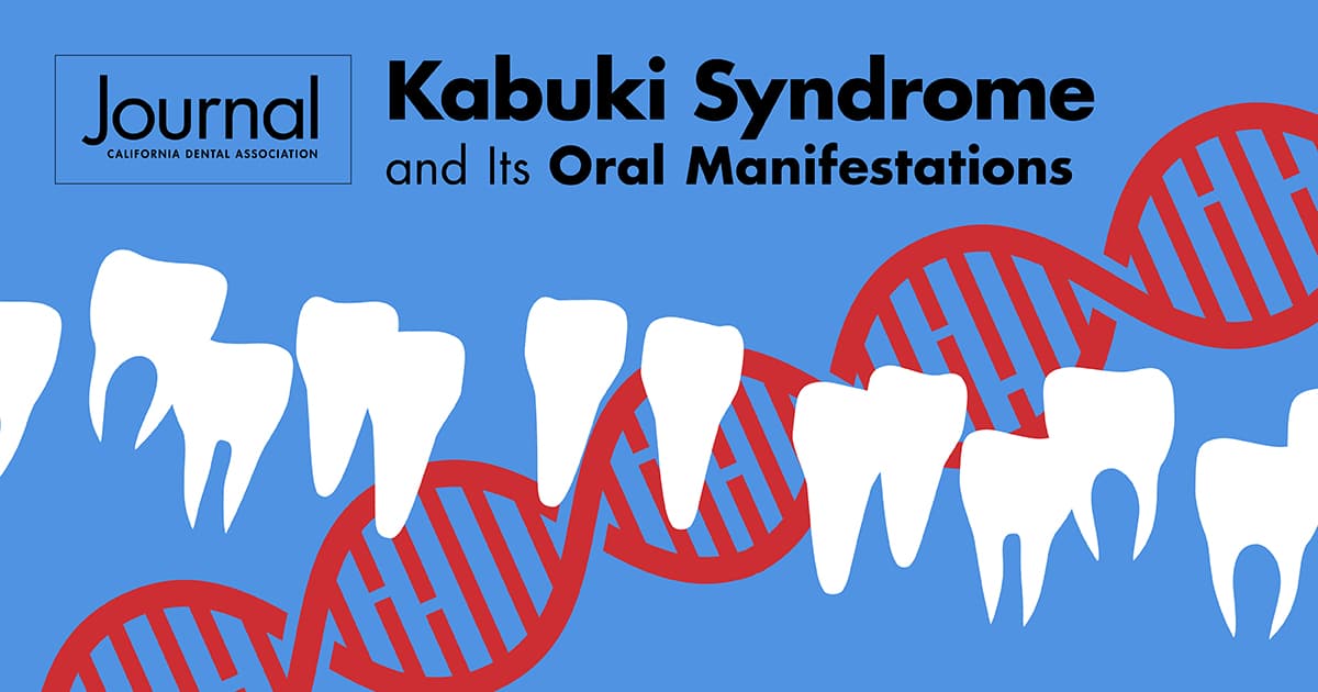
Kabuki Syndrome and Its Oral Manifestations: A Case Report highlights the main oral manifestations of Kabuki syndrome in a 9-year-old patient.
Although Kabuki syndrome is rare and has an etiology that is not yet fully understood, the authors conclude that a diagnosis is not difficult because facial and oral alterations are characteristics of the syndrome.
“The role of the dental surgeon in multidisciplinary teams is highlighted for the correct identification of cases since the facial and intraoral examinations are decisive for the conclusion of the diagnosis,” they write.
Dentists can read the article and successfully complete an online quiz to earn .5 units C.E.
CBCT imaging in diagnosis of craniofacial anomalies
Apert syndrome, achondroplasia, DiGeorge syndrome, Gardner syndrome and several other craniofacial syndromes affect children worldwide with a 2% prevalence rate.
In The Benefits of CBCT Imaging in the Diagnosis of Individuals with Craniofacial Anomalies, the authors discuss the skeletal features and dental abnormalities associated with seven craniofacial syndromes and review the importance of using three-dimensional cone-beam computed tomography to diagnosis the syndromes.
Visualizing structures in three dimensions helps clinicians and radiologists “diagnose and formulate a treatment plan for a multidisciplinary approach for management,” the authors write. “Early diagnosis, and subsequently timely management, is crucial.”
Stem cell therapy in cleft lip and palate
Also included in this collection:
- The implication of Stem Cell Therapy in Cleft Lip and Palate and Other Craniofacial Anomalies — A Literature Review
- Internal Morphological Variations of 150 Lower Premolars: Three-Dimensional Multianalytical Study
- Patient Compliance with Removable Orthodontic Retainers During Retention Phase: A Systematic Review
- Raise Cybersecurity Awareness Among Dental Staff

